- A-
- A+

WHAT IS DEMODEX?
Demodex is an eight-legged mite from the Demodicidae family. There are many species of Demodex but only two of them are found present in humans, demodex folliculorum and demodex brevis.
Demodex mites are found naturally on the faces of human beings from childhood, but especially after the age of 18. Their presence on the skin increases with age.
They live in or around hair follicles (from where hair grows) and sebaceous glands (glands attached to the hair follicles that secrete sebum, an oily substance which prevents the skin and hair from drying out)1.
They form part of what is called the cutaneous microbiota or cutaneous microbial flora, in other words the micro-organisms that naturally live on our skin.

EVOLUTION
Demodex mites need humans in order to live, they develop naturally on our skin until their death2.
The life cycle from the egg to the adult mite lasts between 14 and 18 days. The eggs are deposited in the sebaceous glands and hair follicles, they then turn into larvae until they reach adult form.
In some cases, larger numbers of Demodex develop which is thought to lead to substantial inflammation and result in the appearance of skin lesions. The mechanism behind these reactions, however, is not fully understood.
WHAT ARE THE SYMPTOMS OF DEMODEX?
Demodex mites are not considered to be actual carriers of disease.
However, these parasites may generate conditions that favour the appearance of certain symptoms and pathologies in cases where larger numbers develop on the skin.
During their lifetime, they accumulate waste in their abdomens, which is only released at their death.
In cases where they become too prevalent, this release of residues can cause skin irritation and redness due to chronic inflammation and the development of vascular abnormalities3.

DEMODEX ON THE EYELIDS AND BLEPHARITIS
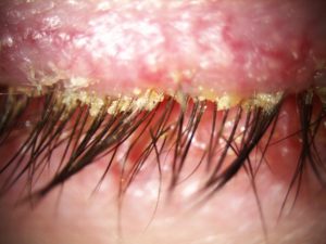
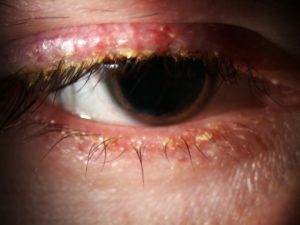
Demodex can be the cause of Blepharitis, which is an inflammation of the edge of the eyelid4.
Demodex Blepharitis manifests itself as non-specific clinical symptoms present in all types of Blepharitis, regardless of the cause (bacteria, viruses, etc.), including:
- redness and itching of the edge of the eyelid;
- grittiness or a foreign body sensation;
- loss of eyelashes;
- and in severe forms, swelling of the eyelids, painful ulcers on the eyelid edges.
It may include some signs such as:
- a pigmentation of the eyelid edge;
- the presence of transparent or whitish flakes forming crusty debris over approximately 1 mm of the base of the lashes: this is the most characteristic and frequent sign and referred to as cylindrical dandruffs 5.
The diagnosis of Demodex Blepharitis is confirmed by the microscopic analysis of an eyelash sample or by visualisation of the parasites with a slit lamp (40-fold magnification) during a consultation with an ophthalmologist.
(1) Action mechanism Théalose brochure
(2) Matsuo T. Trehalose protects corneal epithelial cells from death by drying. The British Journal of Ophthalmology 2001; 5: 610-2.
(3) Cejkova J, Ardan T, Cejka C, et al. Favorable effects of trehalose on the development of UVB-mediated antioxidant/pro-oxidant imbalance in the corneal epithelium, proinflammatory cytokine and matrix metalloproteinase induction, and heat shock protein 70 expression. Graefes Arch Clinical Exp Ophthalmol 2011; 8: 1185-94.
(4) Li J, Roubeix C, Wang Y, et al. Therapeutic efficacy of trehalose eye drops for treatment of murine dry eye induced by an intelligently controlled environmental system. Molecular Vision 2012; 18: 317-29.
(5) Cejkova J, Cejka C, Luyckx J. Trehalose treatment accelerates the healing of UVB-irradiated corneas. Comparative immunohistochemical studies on corneal cryostat sections and corneal impression cytology. Histology and Histopathology 2012; 8: 1029-40.


